
Dr. Roddick’s Remarkable Case of Favus, Showing the Distribution of the Scabs and the Extent of the Eruption on the Body, 1880
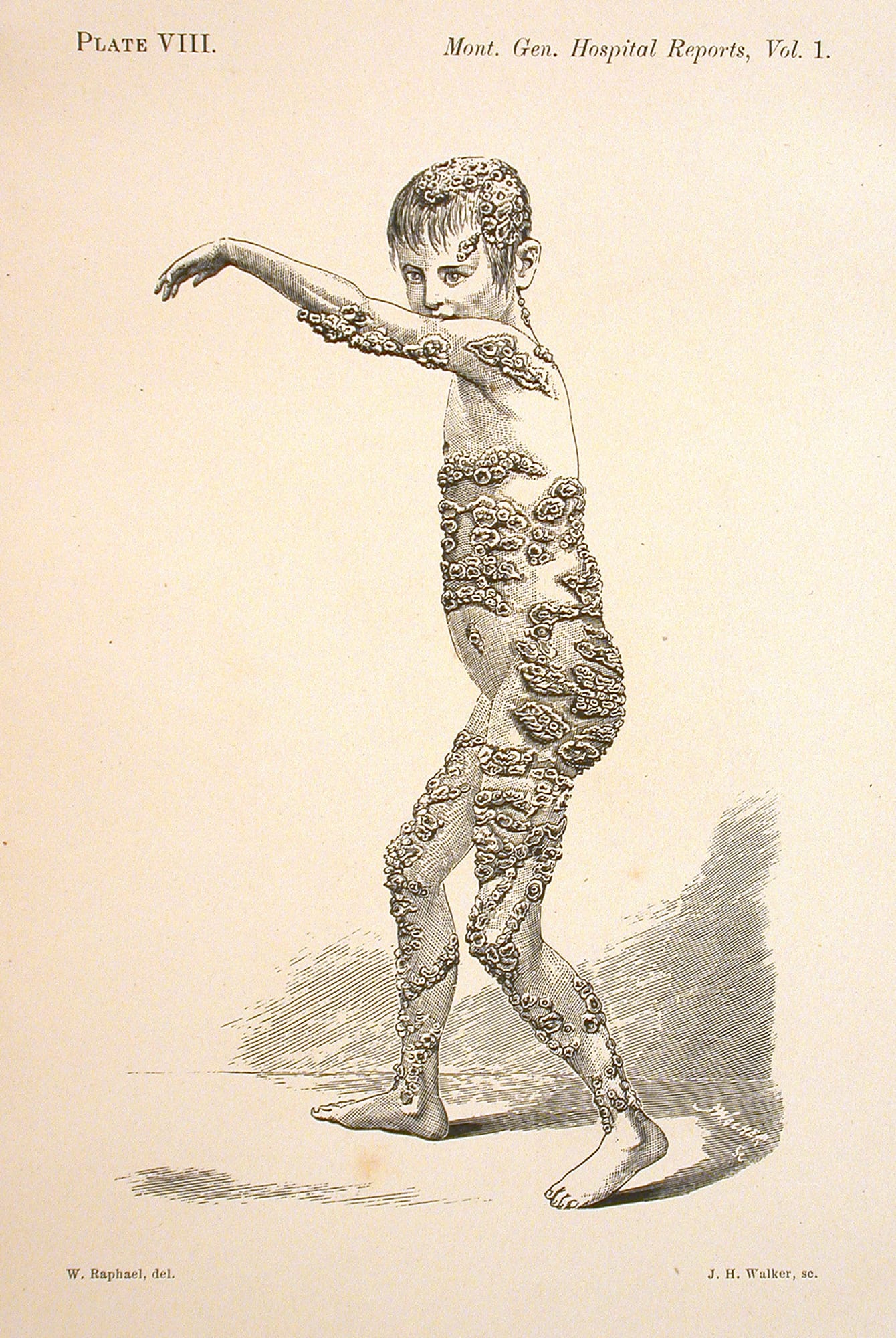
John Henry Walker, after William Raphael, Dr. Roddick’s Remarkable Case of Favus, Showing the Distribution of the Scabs and the Extent of the Eruption on the Body, 1880
Ink on paper, 23.9 x 14 cm
McCord Stewart Museum, Montreal
During the 1870s and 1880s, William Raphael received various commissions from a young physician named William Osler, who had become a professor at McGill University in 1875 and would go on to be one of the most important figures in Canadian medical history. These commissions—drawings of pathologies intended to illustrate the doctor’s research and writings—were nothing like Raphael’s usual subjects. Dr. Roddick’s Remarkable Case of Favus, Showing the Distribution of the Scabs and the Extent of the Eruption on the Body exemplifies his rigour in anatomical representation.
To create these images, Raphael relied as much on a long artistic tradition of anatomical study as he did the collaboration of medical experts. Indeed, during his training in Berlin, he had already acquired detailed knowledge of the human body. The academic approach required students to draw the particularities of the flesh, as well as meticulously examine the body’s interior organs and structures. It was even customary to draw skinned cadavers so as to be able to recognize the various muscles and better capture their details.
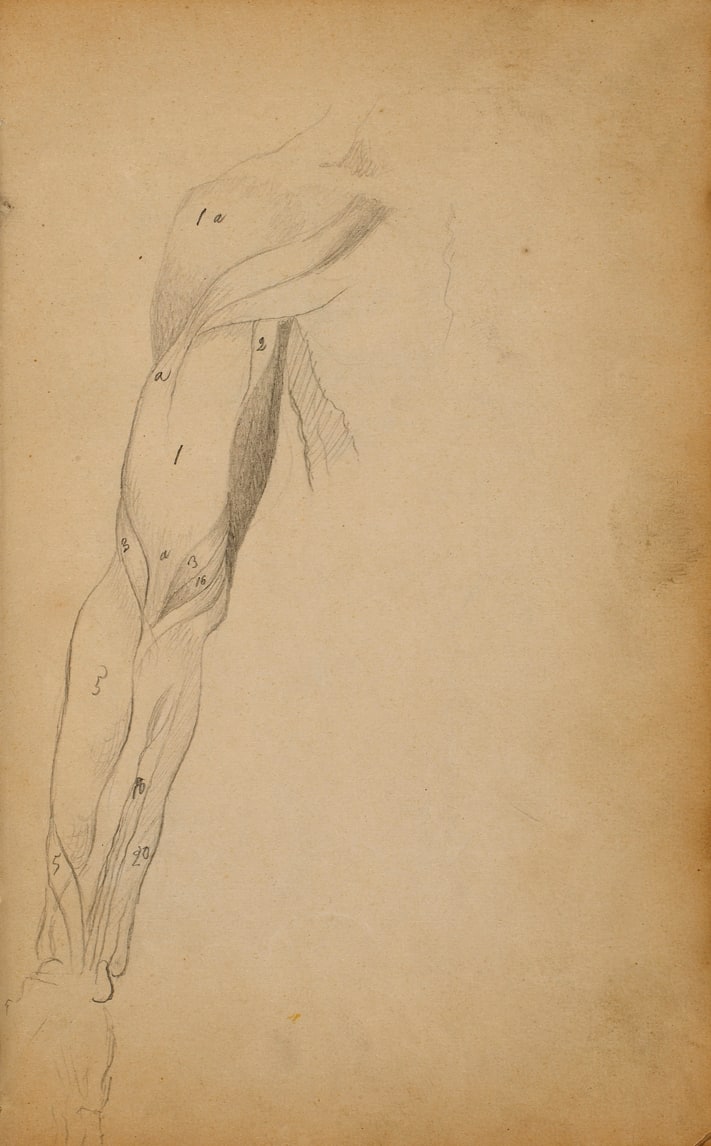

These journeys into medical illustration were also reflected in sketches from Raphael’s student years, when he sometimes drew fragments of flayed bodies. When Osler called upon his talents, Raphael was probably not entirely taken aback by the types of images the doctor wanted. Although the commission was far removed from the quest for beauty that preoccupies many artists, and contact with death and disease can obviously trouble anyone, Raphael proved to be skilled at anatomical draughtsmanship. He was careful to spare no detail, while taking the most objective, dispassionate approach possible.
Osler’s appreciation of Raphael’s work was so great that the doctor even called on the artist from abroad. In 1884, Osler became a professor of medicine at the University of Pennsylvania and began to entrust Raphael with tasks from a distance, noting in one letter that “there is nobody here who can do such fine anatomical work.”
Raphael’s commissions for Dr. Osler included three plates that appeared in the book Montreal General Hospital: Reports Clinical and Pathological, published in 1880 by Dawson Brothers, a well-known local firm. In this book, edited by Osler, Raphael’s work was reproduced by John Henry Walker (1831–1899), an engraver renowned for his preference for xylography (woodcut printing) and his numerous contributions to Montreal newspapers. Plate III features two figures illustrating “Atresia of pulmonary artery, with patent Ductus Arteriosus” and “Ductus Arteriosus and Arch of Aorta in case of General Dropsy of Foetus.” Plate VII features two figures associated with “Dr. Roddick’s Case of Occipital Meningocele.” And plate VIII is the image above, showing a child suffering from an infectious skin disease called favus, a type of ringworm.
The posture of the child, who is seen naked and from the side, reveals the extent of the disease eating away at his skin. At the same time, the pose, with legs apart and left arm raised, is reminiscent of that expected of live models during workshop sessions. Indeed, it was common practice at the time to simulate body movements during poses, as depicted in a drawing by Raphael created in Berlin, Boy Standing in Profile, 1854. The result is a singular image that, for all its interest, did not find a home in Raphael’s painted work; rather, it rested within the pages of Osler’s book, to be seen only by those eager to develop their medical knowledge.
 About the Author
 More Online Art Books
 Acknowledgements